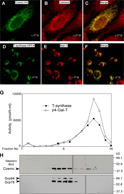
Figure 1.
Localization of human Cosmc and T-synthase. (A–F) Immunofluorescent staining. CHO K1 cells cultured on chambered slides were transiently transfected with Cosmc-HA or with T-synthase–HPC4 and stained with rat anti-HA IgG1 (green) and rabbit anti-calnexin IgG (red; A–C; bars, 4 μm) or with mouse anti-HPC4 (green) and rabbit anti–α-ManII (red; D–F; bars, 8 μm). (G and H) Sucrose gradient subcellular fractionation. 293T cells transiently transfected with HPC4-Cosmc were harvested and homogenized. The postnuclear supernatant (PNS) was loaded onto a sucrose gradient. After ultracentrifugation, 16 fractions (∼0.6 ml/fraction) were collected and measured for both T-synthase and β4–Gal-T activity (G) and analyzed on Western Blot with anti-HPC4 and anti-KDEL (H).

