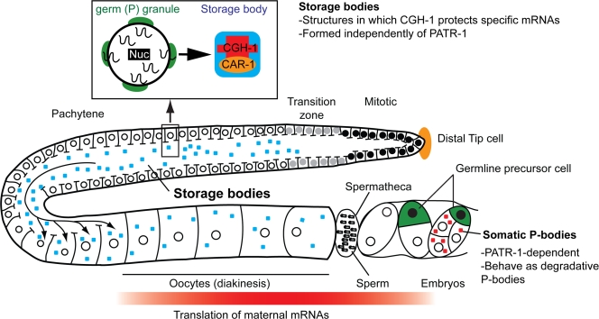
Figure 1.
A C. elegans hermaphrodite gonad, illustrating models discussed in the text. Germ cells develop in an “assembly line” fashion from stem cells that are regulated by the somatic distal tip cell (Hubbard and Greenstein, 2005). Each of two gonad arms produces sperm during the fourth larval stage, then oocytes during adulthood. Germ cells initially share a common cytoplasm, which flows as shown by arrows (Wolke et al., 2007). Transcription increases sharply upon progression through the transition zone and entry into meiosis I. As new mRNAs move into the syncytial gonad core, some appear to pass through germ (P) granules (inset, green), which are located just outside of most nuclear pores (Pitt et al., 2000; Schisa et al., 2001). CGH-1 associates with and protects particular translationally regulated maternal mRNAs in the context of storage bodies, which may be assembled on germ (P) granules (inset). Oocytes enlarge and become cellularized in the proximal region, and fertilization occurs in the spermatheca. Transcription ceases as oocytes enter diakinesis (Walker et al., 2007). Further development depends upon translationally regulated maternal gene products until embryonic transcription begins at the four-cell stage (Seydoux and Dunn, 1997). CGH-1 then associates with PATR-1 in patr-1–dependent somatic P bodies (red dots).

