Figure 2.
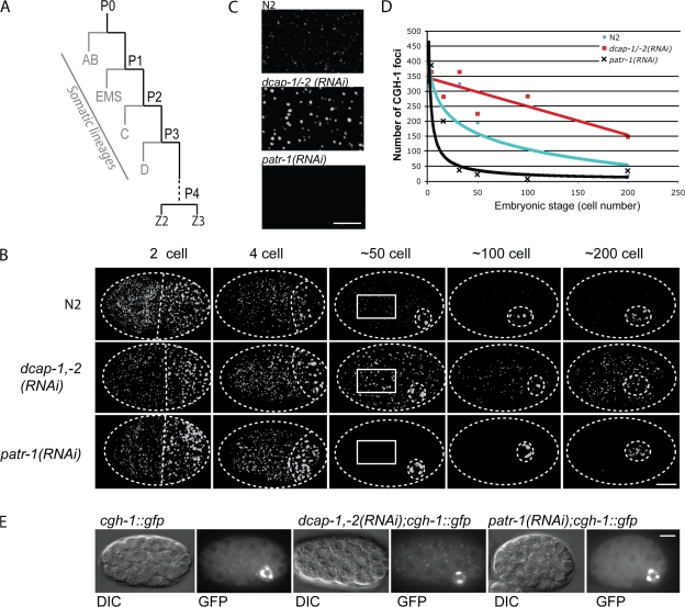
Somatic embryonic CGH-1 foci have characteristics of decapping-associated P bodies. (A) Embryonic founder cells. Four successive asymmetrical divisions of the germ cell precursor each produce a somatic and germ cell daughter. The founding germ line cell P4 gives rise to the germ cell precursors Z2 and Z3 before the 200-cell stage. (B) Effects of dcap-1,-2 and patr-1 RNAi on embryonic CGH-1 foci. Embryos were immunostained for CGH-1 and then analyzed by confocal microscopy. Z series projections of staining throughout the embryo are shown. In germ cell precursors (posteriorly and to the right, outlined by broken lines), most CGH-1 particles colocalize with germ (P) granules (Navarro et al., 2001). At the two- and four-cell stages, CGH-1 foci are distributed similarly in N2 (WT), dcap-1/-2(RNAi), and patr-1(RNAi) embryos. At later stages, in somatic cells, CGH-1 foci are much more prominent in dcap-1/-2(RNAi) embryos than in N2, and are almost absent in patr-1(RNAi) embryos. In each experimental set, >50 embryos were examined at each stage shown. (C) Detailed images of somatic P bodies in 50-cell embryos, corresponding to the boxes in B. (D) Quantitation of somatic embryonic P bodies after dcap-1,-2 or patr-1 RNAi. CGH-1 foci were quantitated in confocal z series projections, with foci of fewer than six pixels and germ (P) granules excluded. Data points each correspond to two-to-seven embryos. (E) Effects of dcap-1,-2 and patr-1 RNAi on embryonic CGH-1∷GFP foci. Single plane differential interference contrast (DIC) and fluorescent images are shown of representative ∼30-cell embryos from a rescuing CGH-1∷GFP transgenic strain. CGH-1∷GFP foci in germ (P) granules were unaffected by these RNAi treatments, but somatic foci were more prominent in decapping-defective embryos than WT, and rarely visible in patr-1(RNAi) embryos. In each set, >40 embryos were examined at this and other early embryonic stages. Bars: (B) 10 μm; (C) 5 μm; (E), 10 μm.

