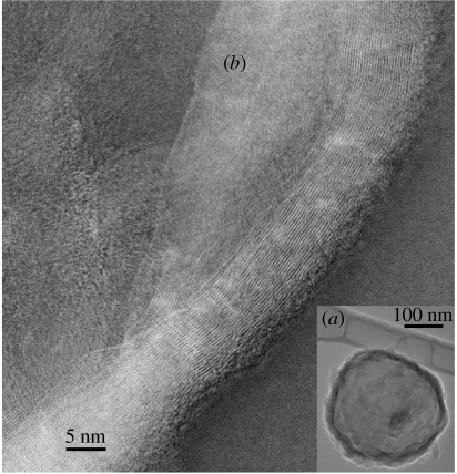
Figure 5.
(a) TEM image of a vesicle consisting of ribbons of subunit c winding around water. (b) TEM inverted image of ribbons of subunit c. A ribbon has β-sheets of strands of subunit c laterally spaced 3.7 Å. The edges of the ribbon away from the vesicle are α-helices. Lengthways the strands/carbon backbones of the protein can be followed in the ribbons as continuous strands of length greater than 134 nm diagonally across the entire image. The nearest ribbon has 24 strands with inter-molecular β-sheet-type bonding and α-helices at its right edge. TEM 200 kV, 1×10⁻⁶ Pa.

