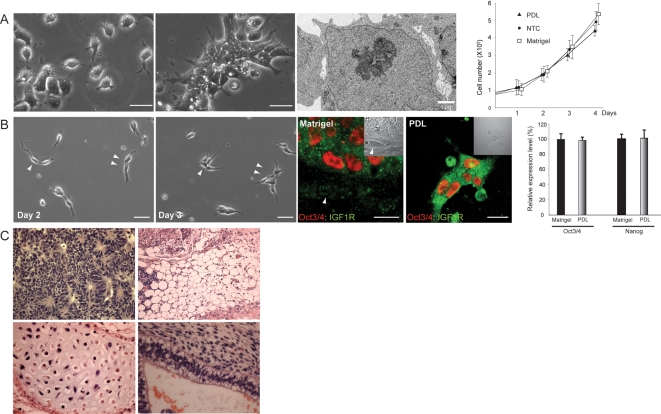
Figure 6. A culture method in which hES Cells self-renew in a completely defined condition.
(A) Morphology of hES cells grown on a poly-D-lysin-coated (PDL) plate in a defined culture medium, mTeSR, supplemented with Y27632. Under this condition, cells initially grew without cell-cell contacts at a low density, whereas, within a few days, cells spontaneously reorganized cell-cell communications and formed small cell clusters with a greater tendency than that of cells grown on NTC plates. Ultrastructural analysis shows that hES cells grown on the PDL plate have underdeveloped cell-cell junctional structures. Cell growth curve indicates that hES cells cultured on PDL plates grow at a speed comparable to that of cells grown on NTC, and slightly slower than that of cells grown on Matrigel-coated plates within 4 days of the culture period. The population doubling time of cells grown on PDL plate was approximately 34.5–38 hrs. Each data point shows mean±SD, n = 3. (B) Sequential microscopic analysis demonstrates the replication of hES cells grown on a PDL plate in the absence of physically contacting fibroblastic differentiated cells. Arrowheads indicate the same cells in the different culture periods. The expression of IGF1 receptor (IGF1R) and Oct3/4 of cells grown on the PDL plate (PDL) was comparable to that of cells grown under the regular condition (Matrigel) as determined by immunocytochemistry. Oct3/4-negative fibroblastic differentiated cells (arrow) grown on Matrigel is negative for IGF1R. Insets show DIC images. QPCR analysis demonstrates that the expression levels of Oct3/4 and Nanog in cells grown under this condition (PDL) are equivalent to that of cells grown under the regular condition (Matrigel). The expression levels (mean±SD, n = 3) are denoted as relative to the expression of each gene in the control condition. (C) hES cells grown on PDL for multiple passages were subjected to teratoma formation assay followed by histological examination. All three germ layer-derivatives such as neuroepithelium (ectoderm), adipose tissue (mesoderm), cartilage (mesoderm), and gland-like epithelium (endoderm) are confirmed. Scale bars, 25 µm.

