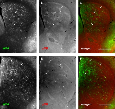
Figure 5. PNN density increases dramatically at postnatal day 14.
Conventions are the same as for Fig 3. (A–C): Rostral sections. (D–F): More caudal sections. (A, D): WFA staining. (B, E): μOR distribution. (C, F): merged. At postnatal day 14, there is a dramatic increase in the number of PNNs: these are found to be distributed selectively within the matrix compartment. There is a marked paucity of proteoglycan expression in the striosome compartment (arrows). A high intensity of diffuse WFA staining associated with the extracellular matrix can be observed in the medial and dorsomedial portions of the neostriatum. Scale bar: 600 μm.

