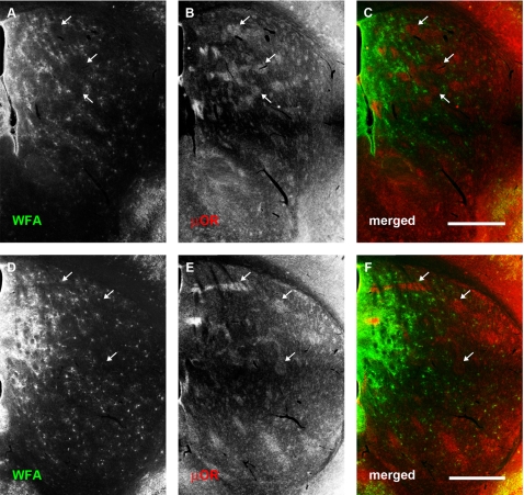
Figure 9. Pattern of WFA staining within the neostriatum continues into adulthood.
Conventions are the same as for Fig 3. (A–C): Rostral sections. (D–F): More caudal sections. (A, D): WFA staining. (B, E): μ-opioid receptor distribution. (C, F): merged. Selective expression of PNNs in the matrix, as well as higher WFA staining in medial extracellular matrix continues into adulthood. Arrows indicate PNN-poor striosomes. Scale bar: 600 μm.

