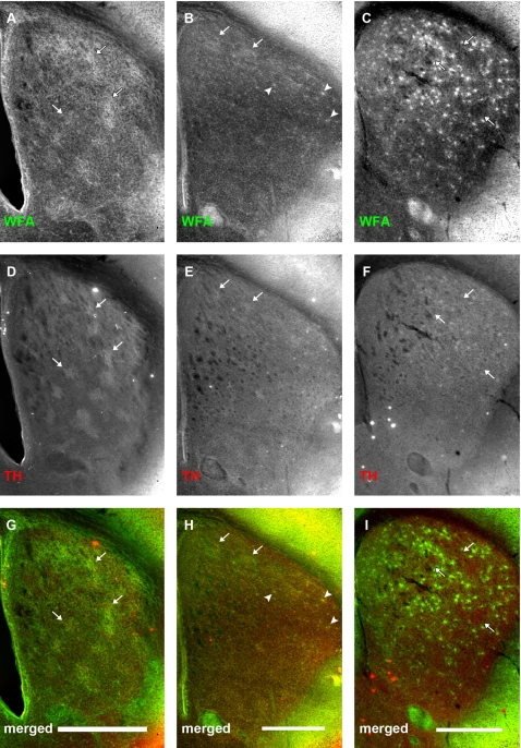
Figure 10. Early association between TH-positive striatonigral terminals and WFA staining dissipates after postnatal day 14.
Panels show relationship between WFA binding (A–C) and tyrosine hydroxylase (TH) expression (D–F) at P4 (A, D, G), P10 (B, E, H) and P14 (C, F, I). (G, H, I) are merged channel images of the two corresponding images above. At P4, TH staining is seen in discrete patches. WFA clusters overlap well with these regions (A, D, G, arrows). At this age, TH-positive dopamine terminals correspond well with μOR-positive striosomes. By P10, TH staining has become considerably more uniform, especially in the medial regions of the neostriatum, than at P4, although some patches can still be observed dorsally. These remaining TH patches can be seen to overlap with WFA clusters (B, E, H, arrows). Arrowheads show PNNs starting to form in the dorsolateral striatum (B, H). By P14, TH staining is uniformly distributed in the entire neostriatum, when WFA staining is predominantly associated with PNNs in the neostriatal matrix (F). Arrows (F, I) mark WFA-poor areas which are reminiscent of striosomes. Scale bar: 600 μm.

