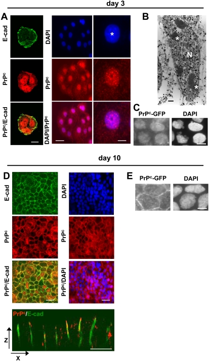
Figure 1. Expression and localization of PrPc in proliferating or differentiated/polarized Caco-2/TC7 cells.
Immunofluorescence labeling of PrPc (red, 12F10 antibody) and E-cadherin (green) was performed after 3 (A, left panel) or 10 (D, left panel) days in culture. Nuclei were stained with DAPI (A, middle and right panels, D, right panel). Right panels in A represent an enlargement of a typical nuclear labeling of one cell (*, nucleolus). Note that left panels correspond to a cluster of three cells and middle panels to a cluster of 13 cells. (B): Immunoelectron microscopy of PrPc was performed on day 3. (N, nucleus; *, nucleolus). (C, E): Sub-cellular localisation of GFP-PrPc was analyzed on day 3 or 10 and compared with DAPI staining (right panels). Note the absence of PrPc and of E-cadherin, used as a marker of the junctional state, at the cell–cell contacts of proliferative cells (A) and their presence in the lateral membranes of polarized/differentiated cells as shown in XY (upper panels D) and XZ (lower panel D) planes. Bars: (A) 10 µm for left panels, 20 µm for middle panels and 4 µm for right panels, (B) 1 µm, (C and E) 10 µm and (D) 20 µm.

