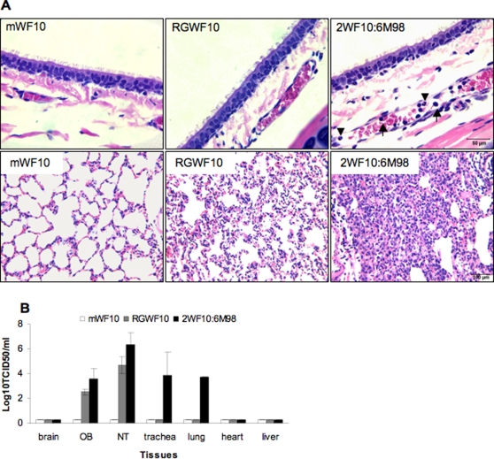
Figure 8. Histopathology and virus distribution of H9N2 viruses in ferrets.
Two ferrets were inoculated i.n. with 106 TCID50 for each virus: mWF10, RGWF10 or 2WF10:6M98. At day 4 p.i, ferrets were euthanized and the tracheas and lungs were harvested for histological analysis. (A) Histopathological findings in the respiratory tract. Upper panel, tracheas: note the margination of neutrophils (▾) and mononuclear cells (↑) in a small vein in the 2WF10:6M98-infected trachea. Lower panel, lungs: note the severe inflammatory infiltration in the 2WF10:6M98-infected lung. (B) Tissue tropism in organs collected from ferrets inoculated with mWF10, RGWF10, or 2WF10:6M98 virus. OB, olfactory bulb. NT, nasal turbinate.

