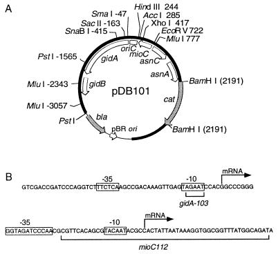
Figure 2.
Structure of oriC plasmid and promoter sequences. (A) The plasmid (pDB101) from which other oriC plasmids are derived is shown with relative restriction sites and genetic map (open arcs). Thick (solid) and thin arcs denote the oriC region and vector sequences, respectively. Shaded arcs denote antibiotic markers. All oriC coordinates are according to refs. 29 and 30. (B) DNA sequences of the gid (7) and mioC (14) promoter regions with promoter mutations are shown. The −10 and −35 consensus homologies and transcriptional start sites are indicated.

