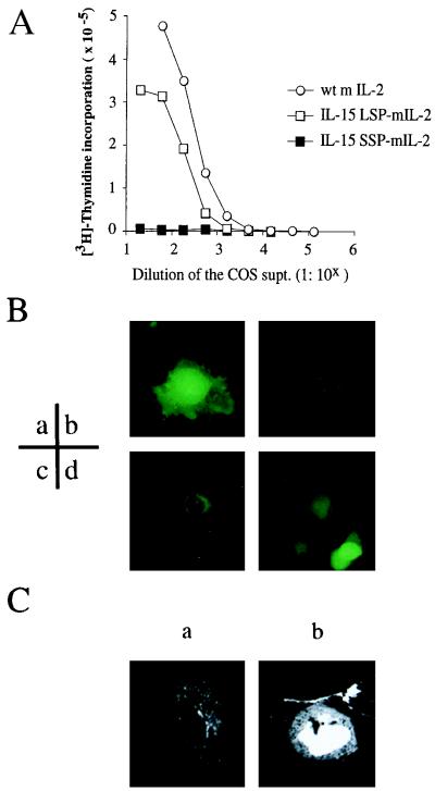
Figure 4.
(A) IL-2 activity detected in the supernatant of COS cells transfected with IL-15 LSP-IL-2, IL-15 SSP-IL-2, or wt IL-2. COS cells transfected the murine IL-2 (mIL-2) constructs with wt IL-2 SP (○), IL-15 LSP (□), or the IL-15 SSP (▪) were cultured for 48 hr. The supernatants of the transfected COS cells were serially diluted and assessed by using the murine CTLL-2 cell line for their secreted IL-2 activity. (B) Immunofluorescence of the COS cells expressing GFP proteins linked with various signal peptides. COS cells transfected with the GFP constructs with various SPs (a, no SP; b, IL-2Rα SP; c, IL-15 LSP; d, IL-15 SSP) were analyzed for their fluorescence by inverted immunofluorescence microscopy. (C) Confocal microscopic analysis of the COS cells expressing the IL-15 LSP-GFP or the IL-15 SSP-GFP protein. COS cells transfected the IL-15 LSP-GFP or the IL-15 SSP-GFP constructs were fixed and analyzed by scanning confocal immunofluorescence microscopy. (a) COS cells transfected with the IL-15 LSP-GFP construct; (b) COS cells transfected with the IL-15 SSP-GFP construct.

