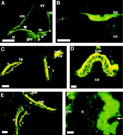
Figure 4.
Otogelin in the inner ear by immunohistofluorescence using the 7.8 antiserum. (A) Cochlea at P0. (B) Cochlea at P15. (C) Vestibule at P0. (D) Anterior crista ampullaris at P4. (E) Saccule and utricle at P20. (F) Saccule at P20 (laser confocal microscopy). The arrowhead in E and F indicates the staining covering the otoconia (○). ger, greater epithelial ridge; M, major tectorial membrane; m, minor tectorial membrane; s, saccule; u, utricle; ca, crista ampullaris; te, transitory epithelium; ne, neuroepithelium; r, roof; cup, cupula; ca, crista ampullaris; om, otoconial membrane; am, accessory membrane. (C) The OMs and the cupula were lost during tissue preparation. Other abbreviations as before. Scale bars: 60 μm (A), 50 μm (B), 100 μm (C–E), 10 μm (F).

