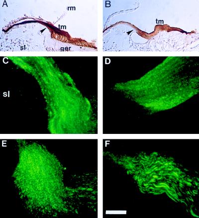
Figure 5.
Otogelin in the tectorial membrane. Immunoperoxidase labeling at P4 (A) and P6 (B) with the 7.8 antiserum. Immunohistofluorescence using the 7.8 antiserum and laser confocal microscopy of the outer edge of the spiral limbus area (arrowhead in A and B) at P4, (C), P6 (D), P8 (E), and P20 (F). The tectorial membrane has the same orientation in all of the figures (A–F). Abbreviations as before. Scale bar: 100 μm (A, B), 10 μm (C–F).

