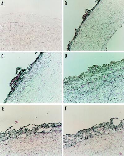
Figure 3.
Localization of NADPH oxidase proteins in rabbit aorta. Aortic sections were incubated with control nonimmune mouse serum (A) or monoclonal antibodies against p22phox (1:100) (B and C), gp91phox (1:200) (D), p47phox (1:20) (E), and p67phox (1:20) (F) at the dilutions shown, followed by gold-conjugated goat anti-mouse antibody. The sections were developed as described in Methods. Magnification is 190× (A and C–F) and 95× (B). The sections shown represent at least three stained sections for each antibody.

