Figure 1.
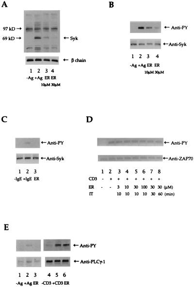
(A) Effect of inhibitors on antigen-induced tyrosine phosphorylation of intracellular proteins in RBL-2H3 cells. Cells (1 × 10⁶ cells per well in 12-well plates) were incubated with 10 μM or 30 μM ER-27319 (ER) for 10 min before stimulation with antigen (Ag; DNP-BSA, 10 ng/ml). Proteins were resolved and transferred to nitrocellulose membranes. The membranes were probed with antibody to phosphotyrosine (Upper) and then reprobed with antibody to FcɛRI β chain (Lower) to normalize for the protein content in each lane. The apparent migration of Syk or FcɛRI β chain is indicated. (B) Effect of ER-27319 on the tyrosine phosphorylation of Syk. Cells (1 × 10⁶) were incubated with 10 μM or 30 μM ER-27319 for 10 min before stimulation with antigen. Cells were lysed and allowed to react with antibody to Syk. Immunoprecipitated proteins was resolved by SDS/10% PAGE and transferred to nitrocellulose membranes. Resolved proteins were probed with antibody to phosphotyrosine (Anti-PY), and subsequently membranes were reprobed with antibody to Syk (Anti-Syk). (C) Immunoprecipitation of Syk from human cultured mast cells. Experimental conditions were identical to those in B but only a concentration of 30 μM ER-27319 was used. (D) Effect of ER-27319 on tyrosine phosphorylation of ZAP-70 in anti-CD3-stimulated Jurkat cells. Cells (1 × 10⁶) were incubated with ER-27319 (at indicated concentrations) for the indicated incubation time (IT) prior to activation with anti-CD3 for 10 min. Cells were lysed and ZAP-70 was immunoprecipitated, resolved, and analyzed by Western blotting with anti-PY and subsequently with anti-ZAP-70. (E) Effect of ER-27319 on tyrosine phosphorylation of PLC-γ1 in antigen-stimulated RBL-2H3 cells or in anti-CD3-stimulated Jurkat T cells. RBL-2H3 cells (4 × 10⁷ cells per 150-cm2 dish) or Jurkat T cells (1 × 10⁷ cells) were incubated with 30 μM ER-27319 (ER) for 10 min before stimulation with 10 ng/ml DNP-BSA (Ag) or 2 μg/ml anti-CD3 (CD3) for 10 min. PLC-γ1 was immunoprecipitated, resolved, and transferred to nitrocellulose membranes. Membranes were probed with anti-PY and subsequently reprobed with anti-PLC-γ1.

