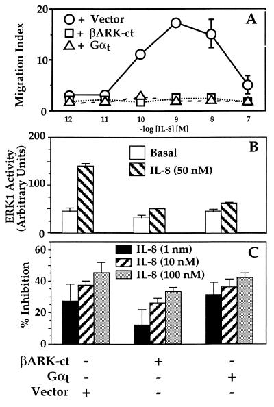
Figure 4.
Responses to IL-8 in cells expressing proteins that sequester βγ. (A) Chemotaxis. Migration assays were performed as described in the legend to Fig. 1 on cells expressing the wild-type IL-8 receptor stably cotransfected with either pcDNA1, pRK-βARK-1-(495–689) (βARK-ct), or αt-pcDNA1, each in combination with a plasmid encoding a hygromycin-resistance marker. (B) MAPK (ERK) activation. Double transfectants were analyzed as described in the legend to Fig. 2. ERK1 activity is expressed in PhosphorImager units. Epidermal growth factor caused 4- to 5-fold activation of ERK in each cell line. (C) Inhibition of cAMP accumulation. Transfectants were incubated with 200 μM forskolin and the indicated concentrations of IL-8. Bars = percent inhibition by IL-8 of the forskolin-stimulated cAMP response. In cells treated with forskolin alone, cAMP accumulation over basal values (measured as the ratio of [3H]cAMP to the sum of [3H]cAMP plus [3H]ATP) was 115, 160, or 247 × 10−3 in cells expressing pcDNA1, βARK-ct, or αt. Results similar to those in each panel were obtained in at least two independent experiments. Data shown represent the mean ± SE of triplicate determinations for A and C and the mean and range of duplicate determinations for B.

