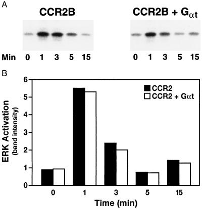
Figure 5.
ERK activation in 300-19 cells expressing CCR2B and Gαt. Stably transfected 300-19 cells were maintained in serum-free medium for 24 h and incubated with 10 nM MCP-1 for the indicated times; lysates were assayed for ERK activation (A) as described. Quantitation is presented in B. Data are representative of three independent experiments.

