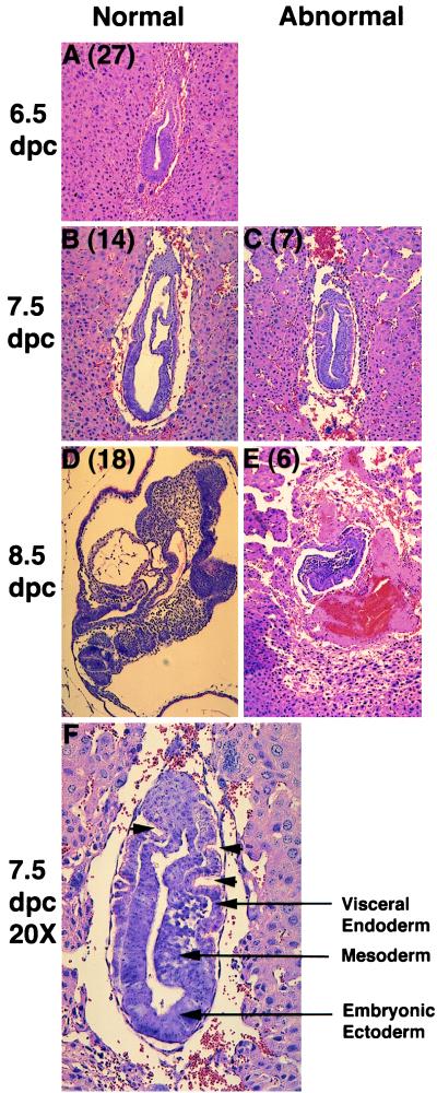
Figure 4.
Histologic analysis of littermate embryos from Dld+/− × Dld+/− matings. The numbers in parentheses indicate the number of embryos with similar phenotypes (A–E, ×10). At 6.5 dpc, all embryos are considered normal because there were no obvious phenotypic differences (A). At subsequent days of development, the littermate embryos could be divided into two phenotypic classes (B–E). A higher magnification of an abnormal embryo at 7.5 dpc is included to allow for more detailed examination (F). Several important cell types are labeled and arrowheads indicate invaginations in the extraembryonic region.

