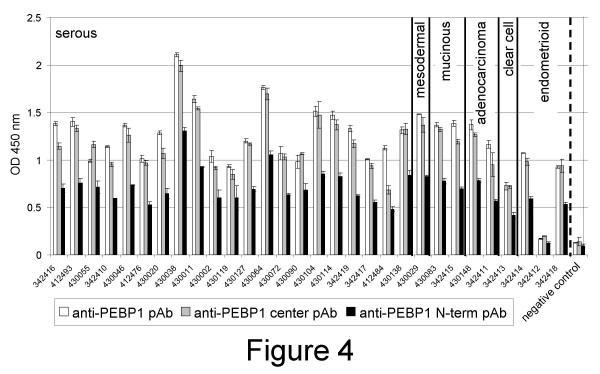
Figure 4.
PEBP1 detection in ascites fluids. Ascites of patients with ovarian cancers from serous mesodermal, mucinous, adenocarcinoma, clear cell or endometrioid origin (as shown) were diluted in binding buffer, coated on plastic wells, and detected with polyclonal antibodies made to the center (pAb, white bars) or N-terminal (pAb, gray bars) region of PEBP1 or with a pAb made to the whole recombinant protein (black bars). Samples were tested in triplicates. Averages are shown. Wells coated with binding buffer were used as negative controls.

