
Figure 5.
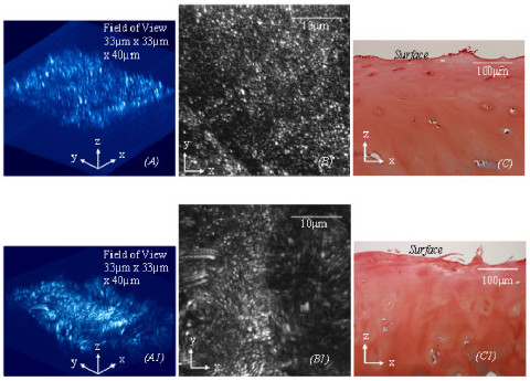
(A). The 3D collagen network (33 μm × 33 um ×) of the cartilage with a matte surface (ICRS Grade 1–2 in Fig 5(C)) obtained from human femoral heads is disrupted and compromised of the collagen fibres aligning predominantly in a direction spatially perpendicular to the AC surface. (A1). The 3D collagen network (33 μm × 33 um) of fibrillated cartilage (ICRS grade 3 in Fig 5(C1)) has an abnormal microstructure and collagen orientation. Images (B)-(B1) are the corresponding MBIs of images (A) and (A1), which are analogous to en face 2D images. Images (C)-(C1) are the corresponding histological images used for ICRS grading.