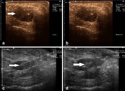
Fig.2.
A 52-year-old female (patient #19) with pain over both the third and fourth metatarsophalangeal joints. A. Longitudinal ultrasound image of the third web space with photopic imaging demonstrates a discrete hypoechoic mass consistent with a neuroma B. Digital calipers indicate the size of the neuroma to be 18.4 × 9.8 mm C. Longitudinal gray scale image demonstrates a linear echogenic needle entering the neuroma for purposes of therapeutic injection (arrow). D. During real-time evaluation, echogenic microbubbles of injected steroid/anesthesia mixture can be seen filling the neuroma (arrow)

