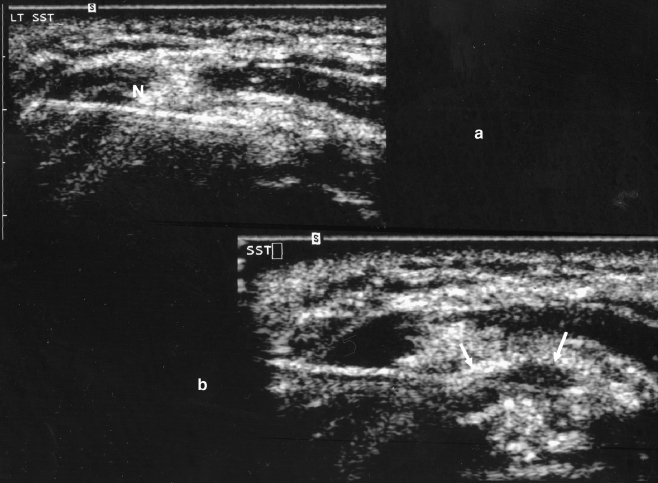
Fig. 2.
a. Ultrasound of the same patient demonstrates needle placement directly into the calcification (N). The tendon is viewed in short axis (rotated 90° relative to its position in Fig. 1). The needle appears linear and echogenic (bright) on ultrasound. b. Ultrasound of the same patient demonstrates the needle, with the tip of the needle demonstrating a smaller area of calcification after mechanical fragmentation and lavage. The boundary of the calcification is depicted by arrows. Fluid replaces some of the echogenic materials after multiple lavages

