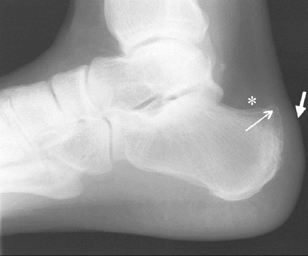
Fig. 1.

Lateral standing radiograph of the hindfoot demonstrates a prominent posterosuperior osseous calcaneal protuberance (arrow) with a vague, cloudy density in the deep retrocalcaneal bursa (*) consistent with retrocalcaneal bursitis. Superficial tendoAchilles bursitis is demonstrated by convexity of the soft tissues (short thick arrow) and an ill-defined Achilles tendon at its insertion consistent with inflammation
