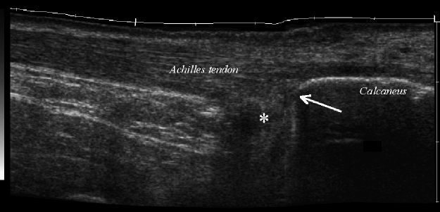
Fig. 2.
Longitudinal extended field of view sonographic image of the hindfoot demonstrates the prominent osseous protuberance at the posterosuperior margin of the calcaneus (arrow) and hypoechoic distention of the retrocalcaneal bursa (*). Thickening of the superficial tendon Achilles bursa is also present

