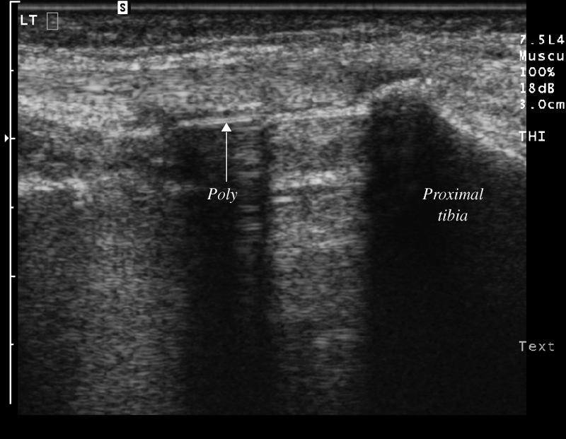
Fig. 4.
Longitudinal ultrasound image along the medial joint line of the knee demonstrating the normal sonographic appearance of a total knee arthroplasty. The polyethylene (arrow, labeled) is seen as a linear echogenic interface with posterior acoustic shadowing. Polyethylene demonstrates similar acoustic characteristics as bone (proximal tibia is labeled). The metallic tibial tray is seen between the polyethylene and the proximal tibia

