Figure 5.
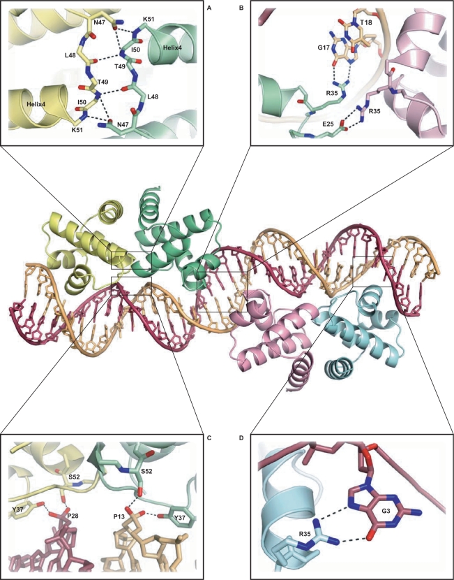
Structure of the C.Esp1396I–DNA complex. Protein subunits are shown in yellow (subunit A), green (subunit B), pink (subunit C) and blue (subunit D). Details are shown of (A) hydrogen bonding at the dimer interface; (B) two alternative R35 contacts from the central subunits B and C, involving both protein–protein and protein–DNA interactions; (C) protein–DNA interactions stabilizing the compressed minor groove around TATA; (D) R35 contacts to the conserved guanine, G3 (outer subunits, A and D).

