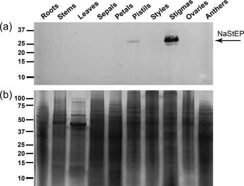
Fig. 3.
Immunoanalysis of NaStEP in N. alata organs. Equal amounts of total protein (10 μg) of roots, stems, leaves, sepals, petals, pistils, styles, stigmas, ovaries, and anthers were separated by SDS–PAGE. (a) Proteins transferred onto nitrocellulose and immunostained with anti-NaStEP antibody. (b) Coomassie blue-stained proteins.

