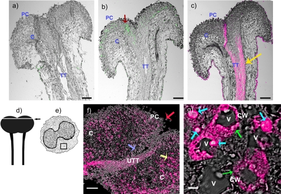
Fig. 6.
Immunohistochemical localization of NaStEP in stigmatic tissues of SC N. alata Breakthrough. (a) Stigma and style treated with pre-immune serum. (b) Stigma and style treated with anti-NaStEP antibody (green). (c) Stigma and style treated with anti-HT-B antibody (magenta). (d) and (e) Diagrams of stigma plus style of N. alata and a transverse section of a stigma. The arrow and square show zones where images were taken. (f) Stigma cross-sections showing NaStEP (magenta) in papillary cells (red arrow), secretory cells (yellow arrow), basal cells (blue arrow), and in a portion of the upper transmitting tract. (g) High magnification view of stigmatic secretory cells shows that NaStEP is in small bodies (blue arrows) and in the proximity of the cell wall (green arrows). All figures represent merges of immunostained and phase contrast images of stigmas plus styles. CW, cell wall; V, vacuole; TT, transmitting tissue; UTT, upper transmitting track; C, cortex; PC, papillae cell. Bars, 50 μm for (a), (b), (c) and 20 μm for (f) and 5 μm for (g).

