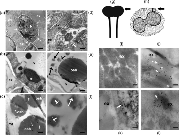
Fig. 8.
NaStEP is discharged from the vacuole to the stigmatic exudate upon pollination. Unpollinated stigma from SC N. alata cv Breakthrough showing NaStEP localization in vacuoles and osmiophilic bodies. (a) Cross-section of mature papillae surrounded by copious exudate containing secretory droplets. Arrows show cell wall interruptions. (b) A papillary cell with a large vacuole and osmiophilic bodies. (c) Anti-NaStEP labelling of vacuolar electron-dense bodies. Arrows show immunogold secondary antibody labelling. Nicotiana alata cv Breakthrough stigma 10 h after pollination with N. tabacum pollen. (d) Degenerating papillary cells (arrow), showing abundant exudate and a disordered cytoplasm with abnormal organelles and disintegrated vacuoles. (e) Vacuole showing poor anti-NaStEP labelling of the osmiophilic bodies (arrows). (f) A close-up of the labelling shown in (e) (black arrows) showing anti-NaStEP labelling (white arrows) in smaller osmiophilic bodies. Stigmatic exudate of N. alata cv Breakthrough. (g) and (h) Diagrams of a stigma plus style of N. alata and a transverse section of a stigma. The arrow and square show zones where images were taken. (i) Unpollinated stigmas. (j) At 10 h after pollination with N. tabacum pollen. (k) At 10 h after pollination with N. plumbaginifolia pollen. (l) At 10 h after self-pollination. N, nucleus; m, mitochondria; c, chloroplast; v, vacuole; osb, osmiophilic bodies; ex, exudate. Bars, 500 nm for (a), 1 μm for (b) and (d), 240 nm for (c), 500 nm for (e), and 200 nm for (i)–(l).

