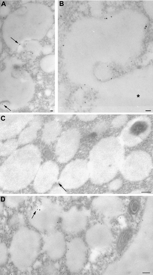
Fig. 2.
Compartments labelled by positively charged nanogold during time-course experiments. (A, B) Labelled MVB fusing with the vacuole (*). The particles were seen in MVBs (see arrows). (C) No nanogold staining was observed in systems of interconnected vesicles forming a single compartment after 15 min of incubation in time-course experiments (arrow: interconnections). (D) Labelling appeared in these compartments after 30 min of incubation with positively charged nanogold and gold particles appeared to detach from the membrane (see arrows). Bar=100 nm.

