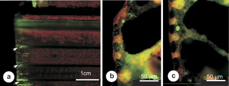
Fig. 3.
Widefield fluorescence micrographs illustrating 5,6-CF transport within cut leaves. (a) The basal 5 cm of the leaf 30 min after placing in 5,6-CFDA solution. Wide distribution of 5,6-CF was seen only in tissues within 1 cm of the severed end of the leaf (arrows). (b) A transverse section of the midrib region, cut in the basal 300 μm of the severed leaf, showing abundant 5,6-CF throughout leaf tissues. (c) Transection of the midrib cut ∼1000 μm from the severed end of the leaf showing 5,6-CF present only within cells of the mesophyll and vascular parenchyma close to the xylem.

