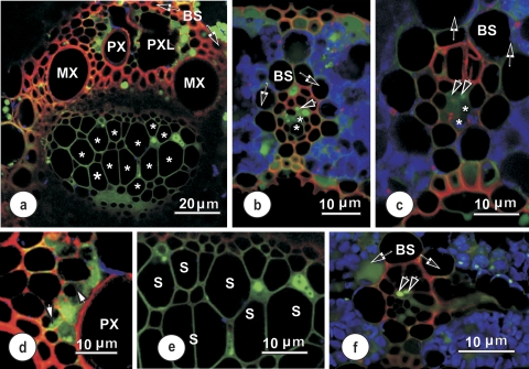
Fig. 5.
Confocal laser scanning micrographs showing the distribution of 5,6-CF (green) and TR (red) in vascular bundles 10–20 cm from the base of a rice leaf, after supplying with 5,6-CDFA together with TR for 60–90 min. Blue coloration indicates chloroplast autofluorescence. (a) Large vascular bundle, showing 5,6-CF and TR distribution. 5,6-CF is localized in parenchyma adjacent to the protoxylem (PX) and some in xylem parenchyma on the phloem side of this vascular bundle. The lumina of large (thin-walled) metaphloem sieve tubes (stars) showed no evidence of 5,6-CF, but the fluorophore appears in all cell walls of the phloem in this section. (b and c) Intermediate longitudinal veins, showing accumulation of 5,6-CF in vascular parenchyma cells. Note: some fluorophore has moved out into mesophyll cells. 5,6-CF is seen in parenchyma elements in the phloem, accumulated in thick-walled (paired arrowheads), but not in the large thin-walled sieve tubes (asterisks). (d) Detail showing the distribution of 5,6-CF in xylem parenchyma adjacent to a protoxylem vessel. Despite some evidence of 5,6-CF in cytoplasm and vacuoles, it was confined to the cytoplasm (arrowheads) one cell removed from the xylem–xylem parenchyma interface. The probe was absent from the protoxylem vessel. (e) Detail of the phloem from (a). Here, 5,6-CF is seen only in phloem parenchyma. Sieve tubes (S) and associated companion cells in this and other undamaged sections contained no 5,6-CF. (f) Transection of a small longitudinal vein, surrounded by a bundle sheath (BS). An emergent thick-walled sieve tube (paired arrowheads point to two TSTs which contain 5,6-CF) in a connecting transverse vein is visible to the right. The TSTs connect to a thick-walled sieve tube exiting from the longitudinal bundle to the right.

