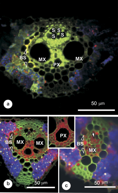
Fig. 6.
Confocal laser scanning micrographs, showing the distribution of LYCH (yellow-green) or 10 kDa TRD (red) in vascular bundles 10–20 cm from the base of a rice leaf, after supplying with either LYCH (a) or TRD (b and c) for 60–90 min. Blue coloration indicates chloroplast autofluorescence. (a) Cross-section of a large vascular bundle showing LYCH in xylem parenchyma between the large metaxylem vessels (MX), in phloem parenchyma, and traces in some large-diameter metaphloem elements (S). Strongly fluorescent patches were seen near the bundle sheath cells (BS), and some fluorescence was seen outside the vascular bundle. (b and c) Distribution of TRD within a large (b) and a small (c) intermediate vein. The xylem parenchyma was very strongly labelled, suggesting rapid offloading to the parenchyma from xylem vessels. TRD spread from the metaxylem (MX) as well as the protoxylem (inset, PX) to xylem parenchyma and phloem-associated parenchyma, as well as thick-walled sieve tubes and some thin-walled sieve tubes (arrow in c).

