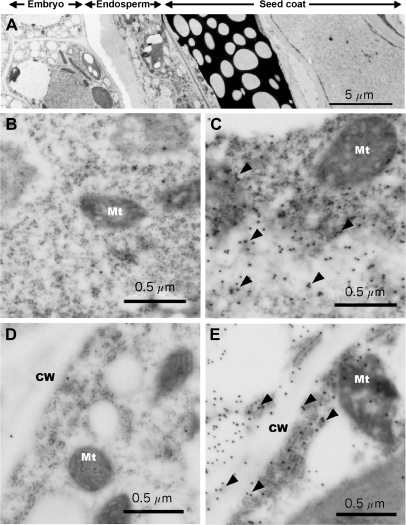
Fig. 2.
Immunoelectron microscopy of MIPS protein in Arabidopsis seeds. (A) Transverse section of whole mature seeds. (B–D) Thin sections of torpedo-stage seeds (B and C) and mature-stage seeds (D and E). B and D show embryo tissue, and C and E show endosperm. CW, cell wall; Mt, mitochondria. Arrowheads show gold particles with anti-AtMIPS2 antibody.

