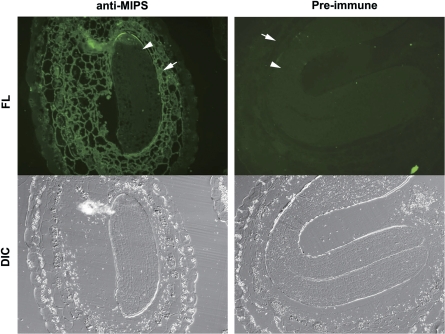
Fig. 3.
Immunofluorescence microscopy of MIPS protein in mature seed sections. Sections were treated with either anti-AtMIPS2 serum (left) or pre-immune serum (right) followed by a secondary anti-rabbit antibody with a fluorescent conjugate (Alexa Fluor 488). DIC, differential interference contrast images; FL, fluorescence images. The arrowhead shows embryo and the arrow shows endsperm tissue.

