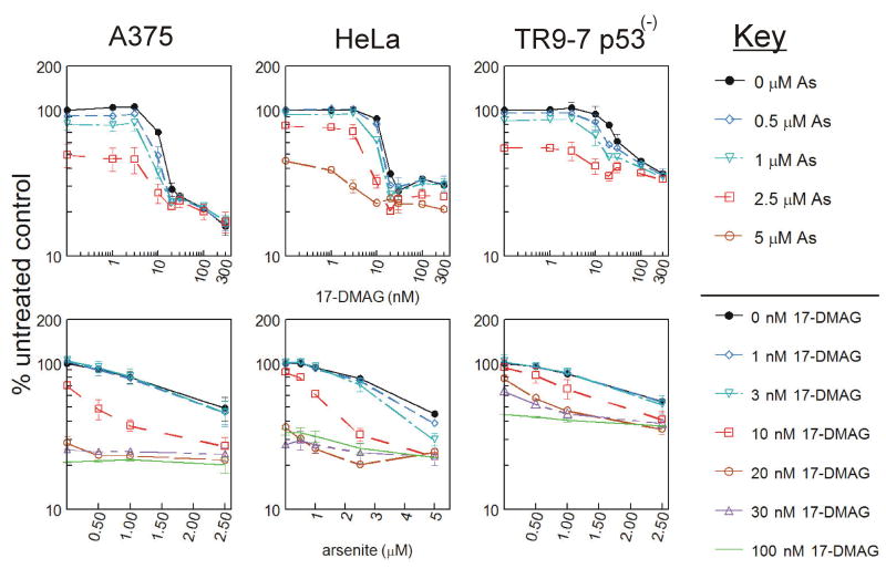
Figure 6. Cytotoxicity of HSP90 inhibitor 17-DMAG co-treatment with arsenite.
A. A375, HeLa, and TR9-7 cells were plated on 96 well dishes in triplicate and exposed to 0-300 nM 17-DMAG in combination with 0, 0.5, 1, 2.5 or 5 μM arsenite for 48 h prior to analysis with AlamarBlue fluorescence assay. Data are graphed as mean ± SD from three independent experiments. Data are presented as percent untreated control either with 17-DMAG concentration on the X-axis (top graphs) or arsenite concentration on the X-axis (bottom graphs).

