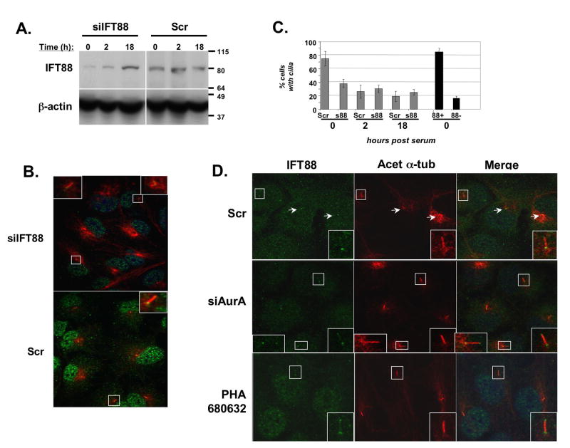
Figure 6. A role for IFT proteins in AurA-induced ciliary resoprtion.
A. Western blot demonstrating siRNA depletion of IFT88 (siIFT88) in ciliated hTERT-RPE1 cells at times following serum treatment, relative to scramble-depleted control. B. Immunofluorescence matching Figure 6A at time 0, indicating relative degree of depletion of IFT88 at the basal body. C. Ciliary disassembly in IFT88- depleted (s88) versus Scr-depleted cells, at 0, 12, or 18 hours after serum treatment, based on the total cell population (gray bars). Black bars (right) indicate % ciliated cells at time 0 calculated specifically from cells confirmed by immunofluorescence to have significant IFT88 staining (88+), or to be well-depleted for IFT88 (88-). D. Cells treated with scrambled (Scr) or AurA-targeting (siAurA) siRNAs, or with PHA-680632 were fixed 2 hours after serum-initiated disassembly. Shown, immunofluorescence indicating cilia (anti-acetylated α-tubulin, red) and IFT88 (green). Insets are enlargements of boxed ciliary structures; arrows indicate direction of ciliary projection relative to basal body. Scale bars, 10 μm.

