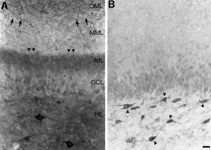
Fig. 1.
Mossy fiber sprouting in slices from pilocarpine-treated rats. A, A section of a hippocampal slice from a rat that developed status epilepticus after pilocarpine treatment illustrates mossy fiber sprouting as a band of neuropeptide Y staining in the inner molecular layer (arrowheads). Note that there also is staining of fibers in the outer molecular layer (small arrows). A few neuropeptide Y-immunoreactive neurons are in the hilus. Scale bar (shown in B): 35 μm. The positions of the layers correspond to both Aand B and are abbreviated as follows:GCL, granule cell layer; HIL, hilus;IML, inner molecular layer; MML, middle molecular layer; OML, outer molecular layer.B, A section from a hippocampal slice of a rat that did not develop mossy fiber sprouting illustrates the normal pattern of neuropeptide Y-like immunoreactivity in the dentate gyrus. Many hilar neurons are stained (arrowheads). Some staining in the outer molecular layer is also normal. There is some staining of granule cells in this section, because it was incubated in DAB for an extended period of time. This was done to increase the likelihood that sprouted fibers would be detectable, if in fact they were present. However, there was no evidence that there was neuropeptide Y-immunoreactive sprouting in this section or in our other control sections.

