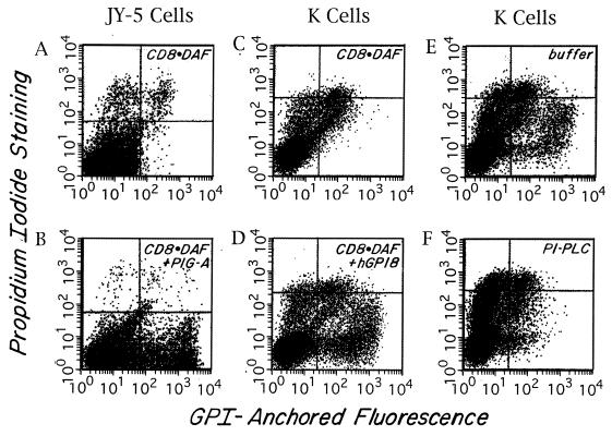
Figure 2.
Rescue of GPI anchoring as assessed in the Vaccinia transient transfection system. Two-color FACS (Becton Dickinson) analyses of anti-CD8 and propidium iodide-stained cells. FL1 (x axis) = CD8 fluorescence intensity; FL2 (y axis) = propidium iodide fluorescence. Cells in the upper quadrants are propidium iodide-positive dead cells. Cells in the lower right quadrant are CD8+ and propidium iodide-negative rescued cells. The small number of cells in the lower right quadrant seen with CD8⋅DAF alone also stained with nonrelevant antibody. (A and B) JY-5 cells transfected with CD8⋅DAF alone or CD8⋅DAF and PIG-A cDNA. (C and D) K cells transfected with CD8⋅DAF alone or CD8⋅DAF and hGPI8. (E and F) CD8⋅DAF and hGPI8 transfected transients treated with buffer or PI-PLC. About 70% of the CD8+ cells were cleaved.

