
Figure 1.
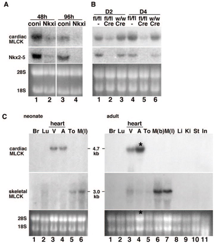
Identification of cardiac-specific MLCK as an Nkx2-5 downstream target gene. A, Northern blotting showed that Nkx2-5 knockdown using adenovirus shRNA-reduced expression of cardiac MLCK 48 hours (lane 1 vs 2) and 96 hours after adenovirus infection (lane 3 vs 4). B, Tamoxifen-induced Nkx2-5 knockout demonstrates reduction of cardiac MLCK expression at postnatal day 2 (D2) (lane 2 vs lanes 1 and 3) and day 4 (D4) (lane 5 vs lanes 4 and 6). Of note, tamoxifen was injected into the pregnant female within 24 hours before delivery. Coni indicates control RNAi; Nkxi, Nkx2-5-RNAi; fl, floxed-Nkx2-5; fl/fl, homozygous for floxed-Nkx2-5; w, wild-type; w/w, homozygous for wild-type; and Cre, Cre-transgene. C, Tissue-specific expression of cardiac MLCK mRNA was examined by Northern blotting (top gels) and compared with that of skeletal MLCK (middle gels) in the neonatal stage (left) and adult stage (right). Increased loading of RNA isolated from adult atrium resulted in higher expression of MLCK in adult atrium vs ventricle (*, lane 4, right). Br indicates brain; Lu, lung; V, ventricle; A, atrium; To, tong; M(l), leg muscle; M(b), back muscle; Li, liver; Ki, kidney; St, stomach; and In, intestine.