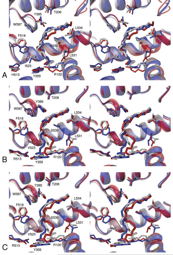
Figure 4.
Stereo views of identical orientations of the COX-1 (A) and COX-2 (B, C) active sites. In each panel, the X-ray crystal structure is shown in gray, the average structure of monomer A in blue and monomer B in red. For COX-2, major (B) and minor (C) average structures are shown, representing two slightly different arachidonate conformations along C11-C15, compared to crystal structure 1CVU, with the productive conformation of arachidonate from the COX-1 crystal structure replacing the inverted arachidonate from the original 1CVU structure.

