
Figure 10.
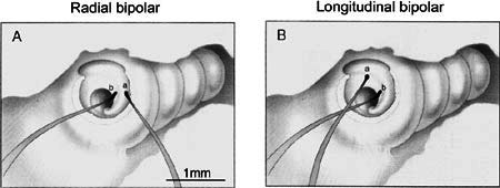
Drawings of VP electrodes in the cochlea of a guinea pig. A. Visually placed ball electrode pair oriented radially at H and 3 on the osseous spiral lamina. B. VP electrodes placed longitudinally spanning the boundary of the hook and first turn. Electrode is placed on the osseous spiral lamina over or just distal to Rosenthal’s canal and the spiral ganglion (location 3, Fig. 8). GP23.