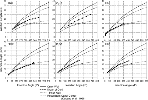
Fig. 3.
Estimation of electrode positions (insertion lengths and angles) for each subject in comparison to the positions of the outer (solid line) and inner (second dashed line) walls of the cochlea, as well as the organ of Corti (first dashed line) and the spiral ganglion (dotted line), as described in the reconstruction work of Kawano et al. (1996).

