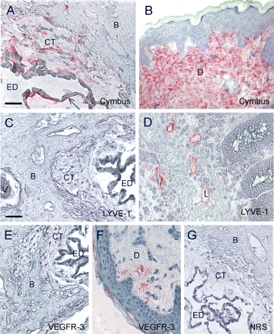
Fig. 2.
Immunohistochemical staining of the human ED using the Cymbus anti-fibroblast antibody (A), staining CT cells especially juxta-positioned to the epithelium (A, arrow), the lymphatic endothelial marker antibodies; anti-LYVE-1 (C) and anti-VEGFR-3 (E). Positive control staining of human skin specimens with the Cymbus antibody (B) and the anti-VEGFR-3 antibody (F), and human large intestine specimen with the anti-LYVE-1 antibody (D). Negative control staining of the human ED with normal rabbit serum (G). Bar 50 μm. ED lumen of the endolymphatic duct, CT interstitial connective tissue, B bone, EP epithelium, L lymph vessel, D dermis, V vein of the VA.

