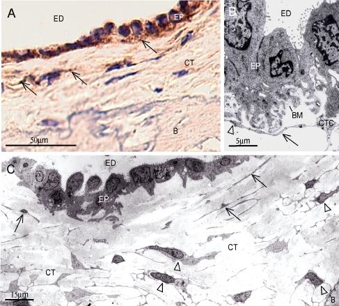
Fig. 3.
A Immunohistochemical staining of the human ED (106) with the anti-platelet derived growth factor receptor β (PDGFR-B2) antibody, showing vesicular staining of CT cells adjacent to the ED epithelium (arrows). Bar 50 μm. B TEM of the human ED epithelium showing epithelia juxta-positioned CT cells. Note long cellular extension in close contact with the basal lamina (arrow) and intercellular contact (arrowhead). Bar 5 μm. C TEM of human ED specimen indicating the presence of a thin lamellar cell type with long cellular extensions located close to the epithelium (arrows), and another more abundant large CT cell type (arrowheads) with numerous cytoplasmatic extensions forming frequent intercellular contacts. Bar 15 μm. ED lumen of the endolymphatic duct, CT interstitial connective tissue, CTC connective tissue cell, B bone, EP epithelium, BM basal membrane.

