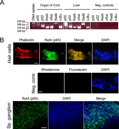
Fig. 1.
NF-κB/Rel expression in the cochlea. (A) RT-PCR showing expression of NF-κB/Rel mRNA in p5 rat organs of Corti and liver (positive control). Negative control reactions were amplified without the RT cycle preceding PCR. (B) Tissue sections of rat p5 cochlea showing localization of RelA (p65) in the hair cells of the organ of Corti (upper panel) and spiral ganglion neurons (lower panel). Phalloidin-rhodamine was used to identify hair cells in the upper panel. Cell nuclei were stained with DAPI (in the lower panel, the condensed nuclei of Schwann cells are stained more intensely than the neuronal nuclei). In the negative controls (middle panel), samples were incubated in absence of RelA (p65) and phalloidin antibodies, but with antirabbit-IgG conjugated to fluorescein. Scale bars represent 15 μm.

