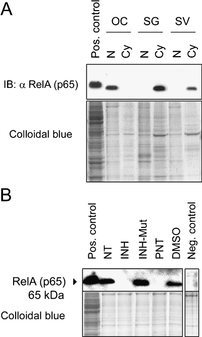
Fig. 2.
Subcellular distribution of NF-κB in the cochlea. (A) Immunoblotting (IB) showing nuclear vs. cytoplasmic distribution of RelA (p65) in the organ of Corti (OC), spiral ganglion (SG), and stria vascularis (SV) lysates; nuclear lysate (N), cytoplasmic lysate (Cy). (B) IB of nuclear lysates prepared from the organ of Corti after treatment with 25 μg/ml inhibitor of NF-κB (INH), 25 μg/ml control for the inhibitor of NF-κB (INH-Mut), 50 mM parthenolide (PNT), or 50 mM DMSO, all for 24 h. In the negative control, nontreated (NT) nuclear protein lysate was incubated with antirabbit-IgG secondary antibody immediately after blocking. All explants were kept in culture for an equal amount of time and collected for protein extraction at the same end time point. Positive controls show HEK293 whole cell lysate; total protein loading amount was visualized by staining with colloidal blue in the lower images.

