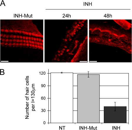
Fig. 3.
NF-κB inhibitors damage hair cells. (A) Middle turns of rat p5 organs of Corti immunostained with phalloidin-rhodamine analyzed by laser scanning confocal microscopy. Representative stacked images obtained with the Multiple Image Processing tool of Imaris are shown. Explants were treated with 25 μg/ml inhibitor of NF-κB (INH) for 24 and 48 h or with 25 μg/ml control for the inhibitor of NF-κB (INH-Mut) for 24 h as indicated. Scale bars represent 20 μm. (B) Quantitative analysis of hair cell survival in organ of Corti basal and middle turns; nontreated (NT), treated with 25 μg/ml inhibitor of NF-κB (INH) or control for the inhibitor of NF-κB (INH-Mut) for 24 h. Values on the y-axis represent total number of hair cells (one row of IHCs + three rows of OHCs) counted per length (l) = 130 μm. Error bars represent standard deviation of the mean.

