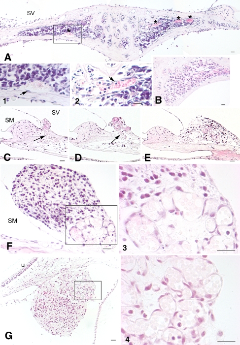
FIG. 6.
Remodeling of blood vessels in early post-injury cochleas. A H&E-stained section through RC taken 3 weeks after ESCs were introduced into RC. Increased blood vessel areas (asterisks) are evident within RC. 1, 2 Higher magnification views of the boxed areas in A showed the morphological characteristics of endothelial cells (arrows) and red blood cells. B Radial section through RC taken from a normal gerbil ear. C–F Sections were taken from another ouabain-exposed cochlea 3 weeks after transplantation. Microvessels (arrows) were found arising from RC across the broken bone and forming a cluster underneath an ESC mass in the supralimbal region. C–E are about 20 μm apart from each other. F is about 40 μm distance from E. 3 Higher magnification of the boxed area in F. G The section was taken from an ouabain-exposed cochlea 3 weeks after transplantation. Association of an ESC mass and a cluster of microvasculature was seen underneath of utricle. 4 Higher magnification of the boxed area in G. Scale bars = 30 μm.

